Symptoms of neurological changes often vary, but may include sensory, motor, behavioural, cognitive or visual symptoms (Shahrokhi & Asuncion 2023).
Neurological observations collect data on a patient’s neurological status and can be used for many reasons, including in order to help with diagnosis, as a baseline observation, following a neurosurgical procedure, and following trauma (CHS 2021).
Therefore, it is important that all healthcare professionals are efficient and accurate in assessing the neurological status of their patients.
It is also important to remember that these changes may occur rapidly over a short period of time or more gradually, taking place over days or weeks. This is why accurate neurological assessments and observations are vital in ensuring the early recognition of neurological deterioration in patients (CHS 2021).
The Neurological Assessment
A neurological assessment involves checking the patient in the main areas in which changes are most likely to occur:
- Level of consciousness
- Pupillary reaction
- Motor function
- Sensory function
- Vital signs.
(QAS 2021a)
Glasgow Coma Scale (GCS)
There are many different assessment tools for neurological function. However, the most widely known and used tool is the Glasgow Coma Scale (GCS).
The patient is assessed and scored in three areas:
- Eye opening
- Verbal response
- Motor response.
(QAS 2021b)
The highest possible score is 15, which reflects an individual who is fully alert, aware and orientated, whereas the lowest possible score is 3, which reflects an unconscious individual.
Although pupil reaction is not included in the GCS, it is often incorporated into the neurological assessment charts used in healthcare facilities in addition to the GCS (Jain et al. 2025).
Because the GCS is widely known, it’s a quick way to communicate a patient’s neurological status and provides a standardised assessment of their neurological functioning. However, if used inconsistently, this can impact its reliability (Jain et al. 2025).
Note that the best response should be recorded for each category (QAS 2021b).
| Behaviour | Rating | Score |
|---|---|---|
| Eye Opening Response | ||
| Opens eyes spontaneously | Spontaneous | 4 |
| Opens eyes in response to speech and sound | Sound | 3 |
| Opens eyes in response to painful stimuli | Pain | 2 |
| Does not open eyes | None | 1 |
| Verbal Response | ||
| Oriented to time, person and place | Oriented | 5 |
| Confused and disoriented | Confused | 4 |
| Utters incoherent words | Words | 3 |
| Incomprehensible sounds | Sounds | 2 |
| Makes no sounds | None | 1 |
| Motor Response | ||
| Obeys two-part requests | Obeys commands | 6 |
| Localises to painful stimuli | Localising | 5 |
| Flexion / withdrawal from painful stimuli | Normal flexion | 4 |
| Abnormal flexion from painful stimuli | Abnormal flexion | 3 |
| Extension to painful stimuli | Extension | 2 |
| Makes no movement | None | 1 |
(Yousuf & Heathcote 2025)
Assessing Level of Consciousness
An individual’s level of consciousness may deteriorate for a number of different reasons, including head injuries, increased intracranial pressure, haemorrhage, lesions or tumours.
Determining a patient’s level of consciousness may be easy or difficult, depending on the individual you are assessing.
In some cases, the patient’s level of consciousness will be obvious when you are talking to them. However, sometimes you may find that they are not responding in an appropriate way or appear confused. Sometimes they may show no response at all.
Sound and physical stimuli are the most common methods for assessing a patient’s level of consciousness (Yousuf & Heathcote 2025).
AVPU Assessment
A rapid assessment tool that is utilised in the healthcare field to measure conscious state is the AVPU scale.
A stands for Alert
The patient is aware of the environment and the examiner and is opening their eyes spontaneously. They can also follow commands and track objects.
V stands for Verbal
The patient’s eyes do not open spontaneously, rather, their eyes only open in response to a verbal stimuli directed towards them. The patient can respond to this verbal stimuli directly and in a meaningful way.
P stands for Pain
The patient's eyes do not open spontaneously or in response to verbal stimuli. The patient will respond to painful stimuli directed towards them by moving, moaning or crying out.
U stands for Unresponsive
The client is not responding spontaneously, or to verbal or painful stimuli.
(Romanelli & Farrell 2023; QAS 2021a)
Assessing Pupillary Reaction
When we assess the patient’s pupils, we gain information about the brain and determine whether there has been an increase in intracranial pressure (Belliveau et al. 2023).
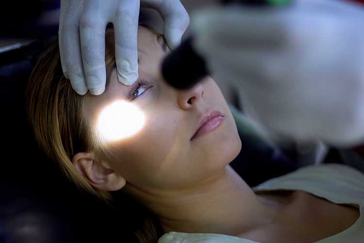
The pupils are assessed for their size and shape, as well as how they react to the presence of light. They should be round and equal in size (Yousuf & Heathcote 2025).
The size of the pupils can vary, but the normal range is 2 to 6 mm in diameter. Upon shining a bright light into each eye, the pupils should constrict briskly to a smaller size (QAS 2021a).
The reactions to light can be described as brisk (normal), sluggish or non-reactive/fixed (Yousuf & Heathcote 2025).
Both eyes should be checked and compared. Generally, any change that occurs during a pupil assessment indicates a change in the individual’s intracranial pressure and may signify a neurological emergency.
Acute pupillary dilation in patients who have suffered a head injury is thought to be caused by compression of the third cranial nerve from brain oedema and herniation (Rauchman et al. 2023).

Assessing Motor Function
Motor function assessment needs to be individualised, and the techniques used should be dependent on the patient’s condition.
For example, if the patient is conscious, the assessment may be made by observing their motor response to commands such as ‘squeeze my hands’. If they are unconscious or unable to provide accurate responses, you may only be able to assess motor function by observation.
You should also note the strength of their limbs, whether they are experiencing any weakness, and whether these effects are bilateral or unilateral.
Limb strength can be described as either:
- Normal power
- Active movement against resistance
- Active movement against gravity
- Active movement with gravity eliminated
- Flicker of movement
- None.
(CHS 2021)
Assessing Vital Signs
Vital signs are also assessed in conjunction with neurological observations in order to gain a full picture of the patient’s current health status.
Changes in vitals may indicate a deterioration of the patient’s neurological condition and can also provide clues to any other medical problems the individual may be experiencing. Depending on the findings of the assessment, further neurological examinations and diagnostic tests may be required.
Overcoming Challenges in Neurological Assessments
There have been concerns raised about the consistency of neurological assessments, especially in regards to assessing pupil size and motor function.
This remains one of the major challenges in regards to neurological assessment. In order to ensure the reliability of neurological assessment and the GCS, it is important that all health professionals conducting these assessments are:
- Fully educated and competent in the use of the GCS and the neurological observation tools being used within their health service, and
- Generating discussion together regarding their patient's neurological condition.
Test Your Knowledge
Question 1 of 3
When assessing pupillary reaction, which one of the following findings is considered normal?
Topics
Further your knowledge
References
- Belliveau, AP, Somani, AN & Dossani, RH 2023, ‘Pupillary Light Reflex’, StatPearls, viewed 22 July 2025, https://www.ncbi.nlm.nih.gov/books/NBK537180/
- Canberra Health Services 2021, Neurological Observation, Australian Capital Territory Government, viewed 22 July 2025, https://www.canberrahealthservices.act.gov.au/about-us/policies-and-guidelines
- Jain, S, Margetis, K & Iverson, LM 2025, ‘Glasgow Coma Scale’, StatPearls, viewed 22 July 2025, https://www.ncbi.nlm.nih.gov/books/NBK513298/
- Queensland Ambulance Service 2021b, Clinical Practice Procedures: Assessment/Glasgow Coma Scale, Queensland Government, viewed 22 July 2025, https://www.ambulance.qld.gov.au/__data/assets/pdf_file/0022/219190/cpp_glasgow-coma-scale.pdf
- Queensland Ambulance Service 2021a, Clinical Practice Procedures: Assessment/Neurological Assessment, Queensland Government, viewed 22 July 2025, https://www.ambulance.qld.gov.au/__data/assets/pdf_file/0027/219195/cpp_neurological-assessment.pdf
- Rauchman, SH, Zubair, A, Jacob, B et al. 2023, ‘Traumatic Brain Injury: Mechanisms, Manifestations, and Visual Sequelae’, Front Neurosci., vol. 17, viewed 22 July 2025, https://pmc.ncbi.nlm.nih.gov/articles/PMC9995859/
- Romanelli, D & Farrell, MW 2023, ‘AVPU Scale’, StatPearls, viewed 22 July 2025, https://www.ncbi.nlm.nih.gov/books/NBK538431/
- Shahrokhi, M & Asuncion, RMD 2023, ‘Neurologic Exam’, StatPearls, viewed 22 July 2025, https://www.ncbi.nlm.nih.gov/books/NBK557589/
- Yousuf, K & Heathcote, T 2025, Glasgow Coma Scale (GCS) and Neurological Observations – OSCE Guide, Geeky Medics, viewed 22 July 2025, https://geekymedics.com/glasgow-coma-scale-gcs/
 New
New 

